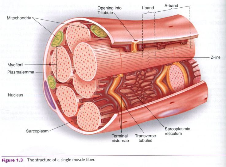
Skeletal muscle fibers can be quite large for human cells with diameters up to 100 μm and lengths up to 30 cm 118 in in the sartorius of the upper leg. Cell Types Skeletal Muscle Cell Atlas Of Plant And Animal Histology -.

The cell membrane is anchored to the cells cytoskeleton by anchor fibers that are approximately 10 nm wide.
Skeletal muscle cell location. Ad Wide Variety of Humanmousecarte Tissue Culture Media and Kits. Normal Human Cells Media High Quality Trusted since 1995. Skeletal muscle cells are also multi-nucleated each cell contains several nuclei.
Some other characteristics of skeletal muscles include. They are attached to bones via tendons. Skeletal muscle tissue can be found across the animal kingdom in most multi-cellular forms of life.
Skeletal muscle is comprised of a series of muscle fibers made of muscle cells. These muscle cells are long and multinucleated. At the ends of each skeletal muscle a tendon connects the muscle to bone.
Skeletal muscle cells a striated muscle cell type form the muscle that we use to move and are compartmentalized into different muscle tissues around the body such as that of the biceps. Skeletal muscles are attached to bones by tendons and can be as long as 30 cm although they are usually 2 to 3 cm in length. Location Of Skeletal Muscle.
Cell Types Skeletal Muscle Cell Atlas Of Plant And Animal Histology -. The functions of skeletal muscles are to bring about specific movements to the number of bones present in the human skeleton according to the university o the functions of skeletal muscles are to bring about specific movements to the numbe. Most Skeletal muscles are found attached to bones by bundles of collagen fibres called tendons.
Skeletal muscles are a form of striated muscle tissue and under the voluntary control of the body. There are approximately 640 skeletal muscles within the human body. They can be categorised into groups relating to.
Frontalis Neck eg sternocleidomastoid Torso eg spinalis Upper. Mononuclear satellite cells are situated between a muscle fiber and its basal lamina. They serve as adult stem cells for the growth and regeneration of the muscle fiber.
Details of both the fine structure and contractile physiology of skeletal muscle fibers are presented in Chapter 5. Skeletal muscle satellite cells are spindle-shaped mononuclear cells located beneath the basal lamina but outside the sarcolemma of skeletal muscle. 74 75 Since their original description by Mauro 76 muscle satellite cells have been primarily identified by their.
Skeletal muscle attaches to the bone by tendons and together they produce all the movements of the body. The skeletal muscle fibers are crossed with a regular pattern of fine red and white lines giving the muscle a distinctive striated appearance. Hence they are also known as striated muscles.
Heart muscle is in the middle of body so heart muscle has nucleus in middle. Skeletal muscles are at periphery of body so nuclei are at periphery. Also you have 1 heart so usually only 1 nucleus per heart muscle cell but have many skeletal muscles so have many nuclei per long fibre.
Skeletal muscle is a fascinating tissue with a complex structure. It consists of elongated multinuclear cells called the myocytes or myofibers. The muscle cells can be anything from 1 mm to 30 cm in length.
The longest muscle cell in our bodies can be found in the sartorius muscle and is 30 cm nearly 12 inches long. Skeletal muscle is a muscle tissue that is attached to the bones and is involved in the functioning of different parts of the body. These muscles are also called voluntary muscles as they come under the control of the nervous system in the body.
Difference between Voluntary and Involuntary Muscles. Skeletal muscle fibers can be quite large compared to other cells with diameters up to 100 μm and lengths up to 30 cm 118 in in the Sartorius of the upper leg. Having many nuclei allows for production of the large amounts of proteins and enzymes needed for maintaining normal function of these large protein dense cells.
Skeletal muscle cells are elongated or tubular. They have multiple nuclei and these nuclei are located on the periphery of the cell. Skeletal muscle is striated.
Smooth muscle cells have a single centrally located nucleus. Cardiac muscle like skeletal muscle is also striated and the cells contain myofibrils myofilaments and sarcomeres as the skeletal muscle cell. The cell membrane is anchored to the cells cytoskeleton by anchor fibers that are approximately 10 nm wide.
These are generally located at the Z lines so that they form grooves and transverse tubules emanate. Because skeletal muscle cells are long and cylindrical they are commonly referred to as muscle fibers. Skeletal muscle fibers can be quite large for human cells with diameters up to 100 μm and lengths up to 30 cm 76 in in the Sartorius of the upper leg.
During early development embryonic myoblasts each with its own nucleus fuse with up to hundreds of other myoblasts to form the multinucleated skeletal. Skeletal muscle fibers can be quite large for human cells with diameters up to 100 μm and lengths up to 30 cm 118 in in the sartorius of the upper leg. During early development embryonic myoblasts each with its own nucleus fuse with up to hundreds of other myoblasts to form the multinucleated skeletal muscle.
Because skeletal muscle cells are long and cylindrical they are commonly referred to as muscle fibers. Skeletal muscle fibers can be quite large for human cells with diameters up to 100 μ m and lengths up to 30 cm 118 in in the Sartorius of the upper leg. The muscle cell or myocyte develops from myoblasts derived from the mesoderm.
Myocytes and their numbers remain relatively constant throughout life. Skeletal muscle tissue is arranged in bundles surrounded by connective tissue. Under the light microscope muscle cells appear striated with many nuclei squeezed along the membranes.
Ad Wide Variety of Humanmousecarte Tissue Culture Media and Kits. Normal Human Cells Media High Quality Trusted since 1995.