Inguinal Ligament bInterosseous membrane between tibia and fibula c. The other two muscles which help in hip flexion are rectus femoris and sartorius.

Lateral and superior surfaces of the greater trochanter of femur.
Muscle that flexes the hip. A muscle in the quadriceps the rectus femoris muscle is attached to the hip and helps to extend or raise the knee. This muscle is also used to flex the thigh. The rectus femoris is the only muscle that can flex the hip.
The hip joint is one of the most flexible joints in the entire human body. The many muscles of the hip provide movement strength and stability to the hip joint and the bones of the hip and thigh. These muscles can be grouped based upon their location and function.
The four groups are the anterior group the posterior group adductor group and finally the abductor group. The anterior muscle group. The pectineus is an accessory hip flexor.
This short muscle originates from the front of the pelvis crosses the hip joint and inserts near the top of the thighbone. In addition to hip flexion the pectineus works with other muscles to move your thigh inward. Hip Flexion Muscles.
There are 11 muscles involved in hip flexion. The amount of activity of each muscle changes depending on how much flexion and whether the femur is in neutral abducted adducted internally rotated or externally rotated. THE MUSCLES INVOLVED ARE.
Psoas Major Iliacus Tensor fasciae latae Sartorius Rectus femoris. The Rectus Femoris muscle is part of the Quadriceps muscle group. It is the only muscle of the group which crosses the hip joint and is a powerful knee extensor when the hip is extended but is weak when the hip is flexed.
Anterior Inferior Iliac Spine AIIS. Top of the patella and the patella tendon to the tibial tuberosity. Pectineus flexes adducts and rotates the thigh at the hip joint as well as stabilizes the pelvis.
Unlike the other muscles in the medial compartment of the thigh it is innervated by the femoral nerve L2-L3 instead of the obturator nerve. What muscles flex the hip. Rectus femoris Tensor fasciae latae Gluteus minimus Sartorius Gluteus medius Psoas major Iliacus Origin and Insertion of Rectus Femoris.
The main flexors of the hip joint are the iliopsoas muscle psoas major and iliacus and the rectus femoris muscle. The pectineus tensor fasciae latae and sartorius muscles assist as weak flexors. The Muscles Crossing the Hip and Knee Joints Anterior View l.
Anterior Medial Pelvic Muscles Iliopsoas. Iliacus and Psoas major. From iliac fossa d.
To lesser trochanter of femur. 2 Psoas major a. The semitendinosus extends the hip and flexes the knee.
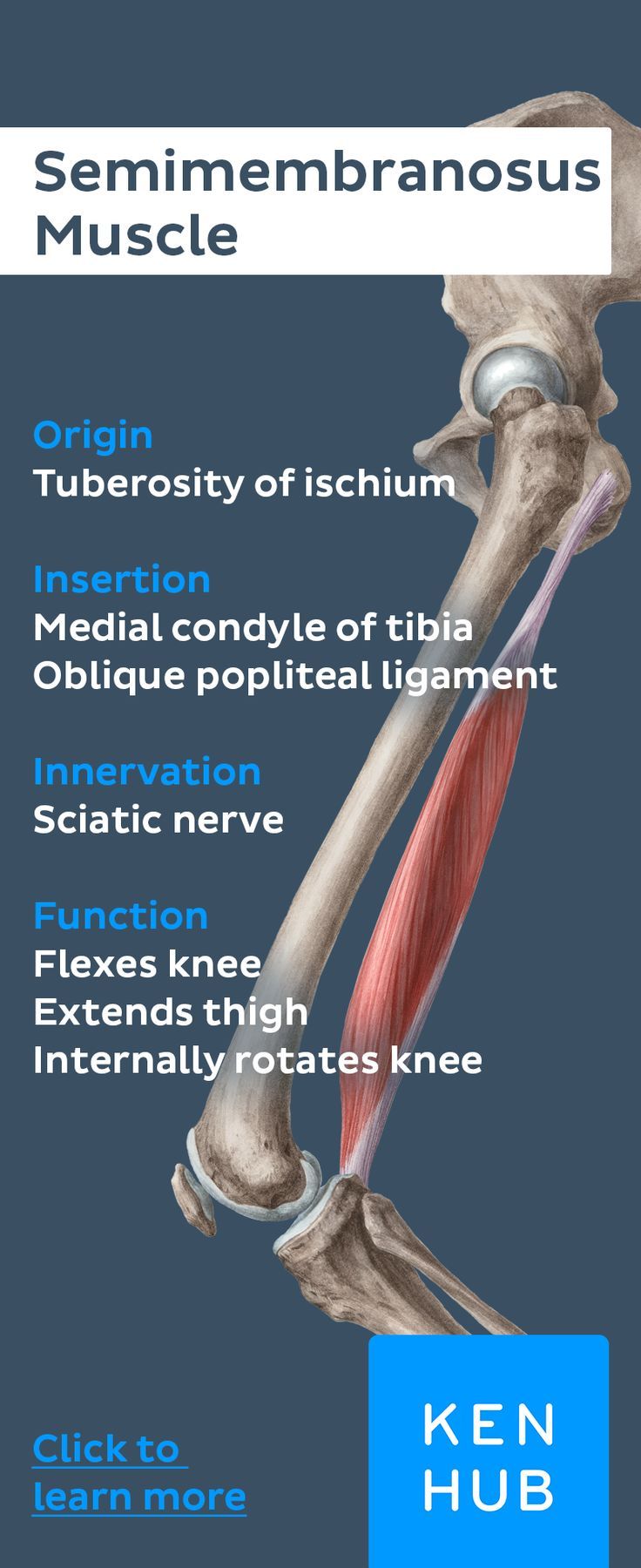
The muscles originate at the ischial tuberosity and insert at the medial surface of the tibial via a structure called the pes anserinus. The semimembranosus extends the thigh flexes the leg and medially rotates the leg when the knee is flexed. The muscle originates on the ischial tuberosity and inserts on the medial condyle of the tibia.
The long head assists in extension of the hip joint. Thigh muscle that causes movement at the. 1- it flexes the leg and rotates it medially.
2- it assists in extension of the hip joint. The hip flexors are in descending order of importance to the action of flexing the hip joint. Collectively known as the iliopsoas or inner hip muscles.
Anterior compartment of thigh. Rectus femoris part of the quadriceps muscle group Sartorius. One of the gluteal muscles.
Medial compartment of thigh. Muscle A flexes internally rotates and abducts the hip Muscle B extends externally rotates and abducts the hip Acting together in a synergy the two muscles can abduct the hip while producing little or no movement in other planes. Contraction of the iliacus and psoas major produces flexion of the hip joint.
When the limb is free to move flexion brings the thigh forward. When the limb is fixed as it is here flexion of both hips brings the body upright. The other two muscles which help in hip flexion are rectus femoris and sartorius.
The following muscle flexes both hip and knee. Pectineus At what defining structure does the Femoral Artery change its name to the Popliteal Artery. Inguinal Ligament bInterosseous membrane between tibia and fibula c.
Femoral triangle The short head of the biceps fempris gets a signal from the nerve. Outer surface of ilium between the posterior and anterior gluteal lines. Lateral and superior surfaces of the greater trochanter of femur.
The fleshy part of the tensor fasciae latae muscle is attached to side of your hip bone. It has a long tendon that extends down to the knee to insert into the tibia which is the larger of your two. Postero-lateral View Sartorius Originates from the Anterior Superior Iliac Spine Inserts onto the medial aspect of upper Tibia Flexes adducts and laterally rotates the hip Flexes the knee 50.
The posterior muscle group is made up of the muscles that extend straighten the thigh at the hip. These muscles include the gluteus maximus muscle the largest muscle in the body and the hamstrings group which consists of the biceps femoris semimembranosus and semitendinosus muscles. Hereof which muscle flexes the thigh at the hip.
These muscle are flexors of the leg at the knee joint but also contribute to extension of the thigh at the hip joint. Sartorius This is a thin strap-like muscle that attaches to the hip bone and crosses the anterior side of the thigh to attach to the medial side of the tibia. The iliopsoas group of muscles iliacus and psoas major is responsible for hip flexion.
The lateral rotator group of muscles externus and internus obturators the piriformis the superior and inferior gemelli and the quadratus femoris turns the anterior surface of the femur outward.