When a nerve impulse arrives at the neuromuscular junction a substance called acetylcholine is released. During contraction the thin filaments slide over the thick filaments.

When the CNS sends a signal the thick and thin myosin filaments form a crossbridge pattern by sliding past each other.
During muscle contraction the. During muscle contraction the myosin heads interact with the thin filament in asynchronous concert in which they appear to walk toward the barbed end of the filament. This is caused by rapid cross-bridge cycling reviewed by Sweeney and Holzbaur 2016. During muscle contraction in humans the A band remains of the same size.
Increase in Ca level into the sarcoplasm leads to the binding of calcium with a subunit of troponin on actin filaments and thereby remove the masking of active sites for myosin. Utilising the energy from ATP hydrolysis the myosin head now binds to the exposed active sites on actin to form a cross bridge. According to sliding-filament theory of muscle contraction the actin and myosin filaments slide past each other with the help of cross-bridge to reduce the length of the sarcomeres.
The smallest unit of muscle contraction is a sarcomere which is delineated by Z- lines. As a muscle contracts the Z lines come closer together. During contraction the thin filaments slide over the thick filaments.
A signal sent by the central nervous system via motor neuron initiates muscle contraction. The neuromuscular junction is the junction between a motor neuron and sarcolemma. A Muscle Contraction Is Triggered When an Action Potential Travels Along the Nerves to the Muscles.
Muscle contraction begins when the nervous system generates a signal. The signal an impulse called an action potential travels through a type of nerve cell called a motor neuron. The neuromuscular junction is the name of the place where the motor neuron reaches a muscle cell.
Skeletal muscle tissue is. During muscular contraction the supply of energy is form the breakdown of ATP. This is broken into adenosine triphosphate ADP and inorganic phosphate Pi and energy is liberated.
Energy liberated by breakdown of ATP is responsible for following activities during muscular contraction. Spread of action potential into the muscle. How much does a muscle shorten during contraction.
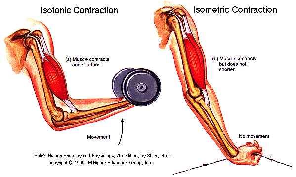
At lengths less than about 70 of resting length the muscle develops no tension at all when stimulated. It follows that during an isotonic contraction a skeletal muscle can only shorten to about 70 of its resting length and it can only develop tension at lengths between 70 and 180 of resting length. During all types of muscular contraction the actin and myosin filaments stay the same length but in isotonic contractions the degree of interdigitation between the two sets of filaments changes as the length of the muscle fibres changes.
The width of the A bands stays the same but the width of the I bands and H zones. The following steps are involved in muscle contraction. 1 The sequence of events leading to contraction is initiated somewhere in the central nervous system either as voluntary activity from the brain or as reflex activity from the spinal cord.
The primary mode of action for muscle is by contraction. What are the steps in muscle contraction. When the CNS sends a signal the thick and thin myosin filaments form a crossbridge pattern by sliding past each other.
This makes the sarcomeres shorter and thicker contracting the muscle. Muscle Contraction Steps in Detail. A signal is sent from the brain or the spinal cord to the muscle via neurons.
Muscle contraction ends when calcium ions are pumped back into the sarcoplasmic reticulum allowing the muscle cell to relax. During stimulation of the muscle cell the motor neuron releases the neurotransmitter acetylcholine which then binds to a. A little muscle contraction fun.
If you prefer a hands on learning experience you might be interested in this giant sarcomere model on Amazon. So lets do a quick review of muscle contraction physiology. An action potential in a motor neuron causes acetylcholine to release in the synaptic cleft.
Acetylcholine binds with receptors on the cell membrane on the muscle fiber opening Ca2 -Na. What moves during muscle contraction. Muscle contraction occurs when sarcomeres shorten as thick and thin filaments slide past each other which is called the sliding filament model of muscle contraction.
ATP provides the energy for cross-bridge formation and filament sliding. During a concentric contraction a muscle is stimulated to contract according to the sliding filament theory. This occurs throughout the length of the muscle generating a force at the origin and insertion causing the muscle to shorten and changing the angle of the joint.
Muscle contraction is brought about by the sliding movement of actin filament over myosin filaments. When a muscle fibril contracts its H-zone disappears A-band remains constant and the I-band shortens while M-line and Z-line overlap. Through the process of muscle contraction tension is developed within muscle tissue which may or may not lead to movement of a part of the body.
The term contraction often means to shorten. However during a muscle contraction the tension may cause muscles. During muscular contraction the myosin heads pull the actin filaments toward one another resulting in a shortened sarcomere.
A sarcomere is defined as the distance between two consecutive Z discs or Z lines. The mechanism of contraction is the binding of myosin to actin forming cross-bridges that generate filament movement Figure 67. This arrangement allows coordinated contraction of the whole muscle in response to neuronal stimulation through a voltage- and calcium-dependent process known as excitationcontraction coupling.
The coupling enables the rapid and coordinated. The sequence of chemical changes that take place during muscle contraction are as follows. Conversion of adenosine triphosphate into adenosine diphosphate.
The first and the important chemical change that takes place during muscle contraction is the conversion of the adenosine triphosphate into the adenosine diphosphate. During muscular contraction the myosin heads pull the actin filaments toward one another resulting in a shortened sarcomere. The I band corresponds to the region of action that does not overlap with myosin.
The length of the actin filament does not change during contraction but the region of overlap increases. Muscle contraction takes place when muscle fibres become shorter. The process of a muscle contraction is split into five stages.
When a nerve impulse arrives at the neuromuscular junction a substance called acetylcholine is released. Muscle contraction allows athletes to apply force and tension during a workout. There are different types of muscle contractions that all help you build strength and mass.
While it might not be something we often think about muscle contraction is used in all kinds of movements and especially in functional fitness.